
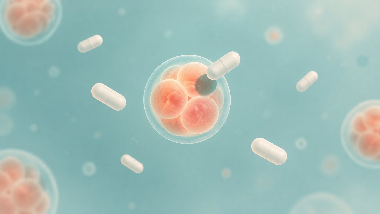
Paracetamol, one of the most widely used painkillers in the world, slows the cell divisions that early embryos depend on, according to a laboratory study from Denmark. After a single standard dose, the drug reached women’s uterine fluid at concentrations that, in culture, delayed or stopped development in human and mouse embryos.
The study was led by Brian S. Nielsen and David M. Kristensen of Copenhagen University Hospital, Rigshospitalet, and Roskilde University, with colleagues from the University of Copenhagen, several Danish fertility clinics and Inserm in Rennes, France. It was published online in Human Reproduction on August 18, 2025.
From yeast to human embryos
The team worked through a chain of models. In fission yeast, paracetamol curbed growth by inhibiting ribonucleotide reductase, an enzyme that makes the building blocks of DNA. Yeast strains with extra enzyme activity were protected. In human kidney cells and two lines of embryonic stem cells, the drug lowered DNA synthesis and caused cells to pile up mid-way through copying their DNA, without killing them.
Next came measurements in women. In 26 fertility patients given 1 g of paracetamol for pain relief, the drug reached an average of 38.1 µM in follicular fluid one hour later. In seven women undergoing a uterine procedure, it averaged 80.3 µM in endometrial tissue and 124.5 µM in uterine fluid, with one woman at 291.3 µM. The uterine level is close to what is typically found in blood after the same dose.
Fewer cells, fewer implantations
Donated surplus IVF embryos were then exposed to 100 or 200 µM. Twenty-two cleavage-stage embryos and 68 blastocysts were used. At 100 µM, early embryos looked structurally normal but had fewer cells. At 200 µM, nuclei fragmented, a sign of cell death. In blastocysts, just six hours of exposure reduced DNA synthesis in the inner cell mass, the cluster that becomes the fetus, and fewer embryos at 100 µM had a clearly definable inner cell mass.
Mouse embryos behaved similarly. At 50 µM and above, two-cell embryos stalled at two to four cells, and none progressed beyond the early blastocyst stage within 48 hours. Exposed embryos transferred into surrogate mice implanted less often and produced fewer live fetuses. Pregnant mice given 200 mg/kg a day by mouth for ten days also had fewer live fetuses and more resorbed embryos.
What the authors caution
The researchers propose three ways the drug could cause loss: outright embryo death at higher doses, failed implantation because development falls behind the womb’s timing, and miscarriage after a weakened inner cell mass. They write that paracetamol “should be used with caution by women attempting to conceive.”
They also flag limits. Only a small number of human cleavage-stage embryos were available, all experiments were in the lab or in animals, and they cannot rule out effects through the drug’s usual pain-relief targets. The mouse feeding dose produced lower levels than those measured in women, because higher doses would damage the liver.
The next step they name is concrete: tracking paracetamol use among healthy women trying to conceive and checking whether it predicts early pregnancy loss.
Study Details:
- Title: Paracetamol (N-acetyl-para-aminophenol) disrupts early embryogenesis by cell cycle inhibition
- Authors: Brian S. Nielsen, Morten R. Petersen, Javier Martin-Gonzalez, Christian Holmberg, Heidi K. Mjoseng, Hanne Frederiksen, Cristal Rosenthal, Emma M. Jørgensen, Palle Serup, Sarah L. Christensen, Kathrine B. Petersen, Karsten Kristiansen, Niklas R. Jørgensen, Jeppe Kari, Anders Hay-Schmidt, Margaux Heurte, Per A. Pedersen, Anders Juul, Anja Pinborg, Søren Ziebe, Svend Lindenberg, Jimmi Elers, Arthur David, Frederikke Lindenberg, Anne Zedeler, Søren T. Christensen, David M. Kristensen
- Journal: Human Reproduction
- Publication Date: August 18, 2025
- DOI: 10.1093/humrep/deaf116